
Nagar Chasmaghar
[/vc_column_text][vc_column_text]Our Expertise in Clinical Support
We are a team of highly skilled optometrists and opticians here to serve your eye care needs. Nagar Chasmaghar was founded in 1957 by brothers, Rameshbhai Ambalal Patel and Jayantibhai Patel. Together they have built an optometric/optical legacy to provide the best eye care services in Gujarat at a budget-friendly price. Today this legacy is being taken forward by the next generation of the family.[/vc_column_text][/vc_column_inner][/vc_row_inner][/vc_column][/vc_row][vc_row disable_element=”yes”][vc_column width=”1/6″][/vc_column][vc_column width=”2/3″][vc_video link=”https://www.youtube.com/watch?v=j6muwUGdvXw” el_width=”60″ align=”center”][/vc_column][vc_column width=”1/6″][/vc_column][/vc_row][vc_section][vc_row][vc_column][vc_separator style=”dotted” border_width=”7″][/vc_column][/vc_row][/vc_section][vc_row][vc_column width=”1/4″][vc_column_text css_animation=”fadeInLeft”]
Concept 01
[/vc_column_text][vc_column_text css_animation=”fadeInLeft”]Zeiss i profiler plus
Benefits
Ocular wavefront and corneal topography
Access to i.Scription® technology by ZEISS
Easy accurate eye measurement
Advanced features for faster workflow with four measurement modalities:
- Ocular wavefront aberrometer
- Autorefractometer,
- ATLAS corneal typographer
- Keratometer
Concept- 02
[/vc_column_text][vc_column_text css_animation=”fadeInLeft”]Retinoscope
Retinoscopy is used to objectively check the refractive state of the eye, especially in kids
Phoropter
Phoropter is used to perform subjective refraction
Concept 03
[/vc_column_text][vc_column_text css_animation=”fadeInLeft”]Portable Autorefractometer
Portable autorefractometer is used for objective refraction during home visits and eye screening camps
[/vc_column_text][/vc_column][vc_column width=”3/4″][vc_masonry_media_grid style=”load-more” items_per_page=”4″ element_width=”3″ btn_style=”modern” btn_color=”default” btn_size=”xs” grid_id=”vc_gid:1715261499766-c414f27a-7886-1″ include=”151840″][/vc_column][/vc_row][vc_row][vc_column][vc_separator style=”dotted” border_width=”7″][/vc_column][/vc_row][vc_row][vc_column width=”1/4″][vc_column_text css_animation=”fadeInLeft”]Concept 04
[/vc_column_text][vc_column_text css_animation=”fadeInLeft”]Non contact tonometer and pachymeter
NCT and pachymeter is used to measure intraocular pressure and thickness of cornea respectively to correlate with any abnormality of eye
[/vc_column_text][/vc_column][vc_column width=”3/4″][vc_masonry_media_grid style=”load-more” items_per_page=”4″ element_width=”3″ btn_style=”modern” btn_color=”default” btn_size=”xs” grid_id=”vc_gid:1715261499767-32e89967-213b-7″ include=”151841″][/vc_column][/vc_row][vc_row][vc_column][vc_separator style=”dotted” border_width=”7″][/vc_column][/vc_row][vc_row][vc_column width=”1/4″][vc_column_text css_animation=”fadeInLeft”]Concept 05
[/vc_column_text][vc_column_text css_animation=”fadeInLeft”]Medmont Corneal topographer
Medmont Corneal topographer is used to map the topography of the cornea for Specialty Contact lens and Scleral lens fitting
[/vc_column_text][/vc_column][vc_column width=”3/4″][vc_masonry_media_grid style=”load-more” items_per_page=”4″ element_width=”3″ btn_style=”modern” btn_color=”default” btn_size=”xs” grid_id=”vc_gid:1715261499767-6ba6b1f4-d66b-4″ include=”151842″][/vc_column][/vc_row][vc_row][vc_column][vc_separator style=”dotted” border_width=”7″][/vc_column][/vc_row][vc_row][vc_column width=”1/4″][vc_column_text css_animation=”fadeInLeft”]Concept 06
[/vc_column_text][vc_column_text css_animation=”fadeInLeft”]Slit Lamp microscope
Slit Lamp microscope is used to examine ocular structures , document ocular conditions such as dry eyes and contact lens fitting
[/vc_column_text][/vc_column][vc_column width=”3/4″][vc_masonry_media_grid style=”load-more” items_per_page=”4″ element_width=”3″ btn_style=”modern” btn_color=”default” btn_size=”xs” grid_id=”vc_gid:1715261499768-51a6ad45-e437-10″ include=”151843″][/vc_column][/vc_row][vc_row][vc_column][vc_separator style=”dotted” border_width=”7″][/vc_column][/vc_row][vc_row][vc_column width=”1/4″][vc_column_text css_animation=”fadeInLeft”]Concept 07
[/vc_column_text][vc_column_text css_animation=”fadeInLeft”]Fundus photographer
Fundus photographer is used to check health of retina and diagnosis of retinal diseases in Hypertensive and Diabetic patients
[/vc_column_text][/vc_column][vc_column width=”3/4″][vc_masonry_media_grid style=”load-more” items_per_page=”4″ element_width=”3″ btn_style=”modern” btn_color=”default” btn_size=”xs” grid_id=”vc_gid:1715261499769-05faeb9e-ab7e-1″ include=”151848″][/vc_column][/vc_row][vc_row][vc_column][vc_separator style=”dotted” border_width=”7″][/vc_column][/vc_row][vc_row][vc_column width=”1/4″][vc_column_text css_animation=”fadeInLeft”]Concept 08
[/vc_column_text][vc_column_text css_animation=”fadeInLeft”]Paediatric Eye Examination
Thorough eye examination including Torch light examination, color vision test, cover test and ocular motility test is performed for paediatric patients
[/vc_column_text][/vc_column][vc_column width=”3/4″][vc_masonry_media_grid style=”load-more” items_per_page=”4″ element_width=”3″ btn_style=”modern” btn_color=”default” btn_size=”xs” grid_id=”vc_gid:1715261499769-c7508089-8003-7″ include=”151849,151850″][/vc_column][/vc_row][vc_row][vc_column][vc_separator style=”dotted” border_width=”7″][/vc_column][/vc_row][vc_row][vc_column width=”1/4″][vc_column_text css_animation=”fadeInLeft”]Concept 09
[/vc_column_text][vc_column_text css_animation=”fadeInLeft”]Non strabismic evaluation and management
Non-strabismic evaluation including worth four dot test, ocular motility test, battery test, etc is performed to rule out accommodative and vergence abnormalities and manage them
[/vc_column_text][/vc_column][vc_column width=”3/4″][vc_masonry_media_grid style=”load-more” items_per_page=”4″ element_width=”3″ btn_style=”modern” btn_color=”default” btn_size=”xs” grid_id=”vc_gid:1715261506896-6eb074f3-d4f5-5″ include=”151932,151933″][/vc_column][/vc_row][vc_row][vc_column][vc_separator style=”dotted” border_width=”7″][/vc_column][/vc_row][vc_row][vc_column width=”1/4″][vc_column_text css_animation=”fadeInLeft”]Concept 10
[/vc_column_text][vc_column_text css_animation=”fadeInLeft”]Orthokeratology & Myopia Control
Myopia control to slow down the progression of myopia in children using specially designed Ophthalmic lenses like ZEISS Myocare, Myokids ,Myovision Pro, Essilor Stellest & Hoya Myosmart lenses.
[/vc_column_text][/vc_column][vc_column width=”3/4″][vc_masonry_media_grid style=”load-more” items_per_page=”4″ element_width=”3″ btn_style=”modern” btn_color=”default” btn_size=”xs” grid_id=”vc_gid:1715261617277-bce3a23a-160c-1″ include=”151936″][/vc_column][/vc_row][vc_row disable_element=”yes”][vc_column width=”1/6″][/vc_column][vc_column width=”2/3″][/vc_column][vc_column width=”1/6″][/vc_column][/vc_row][vc_section][vc_row][vc_column][vc_separator style=”dotted” border_width=”7″][/vc_column][/vc_row][vc_row][vc_column][vc_column_text css_animation=”fadeInLeft”]Human Resources
[/vc_column_text][/vc_column][/vc_row][vc_row][vc_column width=”1/3″][vc_single_image image=”100080″ img_size=”medium” alignment=”center” onclick=”zoom” css_animation=”fadeInUp”][vc_column_text css_animation=”fadeInLeft”]Gauri Patel
M.Optometry, FIACLE (Australia), FBCLA (UK)

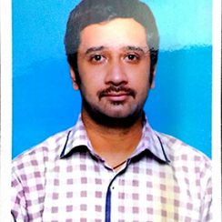
[/vc_column_text][/vc_column][vc_column width=”1/3″][vc_single_image image=”100086″ img_size=”medium” alignment=”center” onclick=”zoom” css_animation=”fadeInUp”][vc_column_text css_animation=”fadeInLeft”]Rushit Patel
M.Optom, FIACLE (Australia)
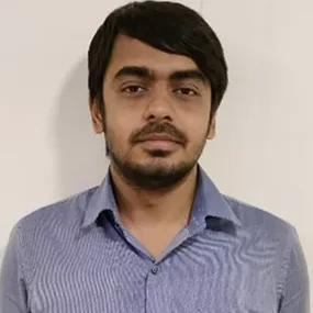
[/vc_column_text][/vc_column][vc_column width=”1/3″][vc_single_image image=”100083″ img_size=”medium” alignment=”center” onclick=”zoom” css_animation=”fadeInUp”][vc_column_text css_animation=”fadeInLeft”]Nihar
B.Sc Optom, FCL (Sankara Netralaya)
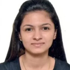
[/vc_column_text][/vc_column][/vc_row][vc_row][vc_column width=”1/3″][vc_single_image image=”107982″ img_size=”medium” alignment=”center” onclick=”zoom” css_animation=”fadeInUp”][vc_column_text css_animation=”fadeInLeft”]Aanal Raja
B.optom, Fellowship (paediatric optometry)
[/vc_column_text][/vc_column][vc_column width=”1/3″][vc_single_image image=”107985″ img_size=”medium” alignment=”center” onclick=”zoom” css_animation=”fadeInUp”][vc_column_text css_animation=”fadeInLeft”]Devanshi Jadav
B.Optom
[/vc_column_text][/vc_column][vc_column width=”1/3″][vc_single_image image=”107988″ img_size=”medium” alignment=”center” onclick=”zoom” css_animation=”fadeInUp”][vc_column_text css_animation=”fadeInLeft”]Simran Ramchandani
M.Optom, FCL (IAO)
[/vc_column_text][/vc_column][/vc_row][/vc_section][vc_section css=”.vc_custom_1561545526154{background-color: #dd9933 !important;}”][vc_row][vc_column][vc_empty_space height=”15″][/vc_column][/vc_row][/vc_section][vc_section][vc_row][vc_column][vc_column_text]