
Mukta Opticians
Founded in 1977 by Sarsolkar Brothers, MUKTA OPTICIANS offers eye wear services which is known for quality at affordable price. The vision to provide best quality eye care service has been the dream and passion of the proprietor Mr. Kishor Sarsolkar and Mukta Opticians have been successful in providing best quality vision possible to the customers. MUKTA OPTICIANS currently serves over a hundred thousand customers in state of Goa and Maharashtra and Karnataka and employs more than 50 people in the greater Goa across its retail outlets in Mapusa, Comba, Curchorem, Margao, Panaji, Sanquelim, Sawantwadi with its head office at Dhargal, Goa.
Equipment
Handheld Ref/Keratometer
The HandyRef-K/HandyRef is lightweight and also has excellent weight distribution. Its compact design makes it easy to hold, balance, and use. Intelligently designed button layout is also useful in one-handed operation.
Equipment
NIDEK Tabletop Refraction Unit
The TS-610 and TS-310 are tabletop subjective refraction workstations that integrate chart and refractor into a single unit. NIDEK’s recognized pedigree of quality examination process is embodied in a groundbreaking design that redefines conventional refraction systems and significantly minimizes the examination footprint. You can choose between two different models, which are the high-end TS-610, offering a more advanced examination, and the standard TS-310 with core examination functions.
Equipment
Automated Refractometer by NIDEK
An Autorefractor or Automated refractor is a computer-controlled machine used during an eye examination to provide an objective measurement of a person’s refractive error and a prescription for glasses or contact lenses. This is achieved by measuring how light is changed as it enters a person’s eye.
Equipment
FUNDUS Camera by 3nethra
The 3nethra classic is a digital non-mydriatic fundus camera, equipped with an efficient workflow to capture undistorted and uniformly illuminated photographs of the retina and surface area of the cornea. The compact design of the camera enhances mobility and easy deployment. The camera is used by clinicians in ophthalmology, optometry, or primary care practice to assist them in the effective evaluation, diagnosis, and documentation of visual health. Some of the common eye diseases that lead to visual impairment are glaucoma, diabetic retinopathy, Age-Related Macular Degeneration (ARMD), and cataract.
Equipment
Direct Ophthalmoscope
Ophthalmoscope an instrument for examining the interior of the eye.
direct ophthalmoscope one that produces an upright, or unreversed, image of approximately 15 times magnification. The direct ophthalmoscope is used to inspect the fundus of the eye, which is the back portion of the interior eyeball. Examination is best carried out in a darkened room. The examiner looks for changes in the color or pigment of the fundus, changes in the caliber and shape of retinal blood vessels, and any abnormalities in the macula lutea, the portion of the retina that receives and analyzes light only from the very center of the visual field. Macular degeneration and opacities of the lens can be seen through direct ophthalmoscopy.
Equipment
Slit Lamp Biomicroscope
A slit-lamp is a binocular microscope used for eye examination using a slit-like light beam. In 1911, Allvar Gullstranda Swedish Ophthalmologist designed table-mounted binocular eyepiece for 3-dimensional visualization of optically clear eye structures. Later Otto Henker, combined Gullstrand slit lamp with Czapski’s binocular microscope resulting in first slit lamp biomicroscope, allowing hand-free examination of an eye.
Equipment
Bscan Ocular Ultrasonography
B-scan ultrasonography is an important adjuvant for the clinical assessment of various ocular and orbital diseases. With understanding of the indications for ultrasonography and proper examination technique, one can gather a vast amount of information not possible with clinical examination alone. This article is designed to describe the principles, techniques, and indications for echographic examination, as well as to provide a general understanding of echographic characteristics of various ocular pathologies.
Equipment
Optical Coherence Tomography
Optical coherence tomography (OCT) is a non-invasive imaging test. OCT uses light waves to take cross-section pictures of your retina. With OCT, your ophthalmologist can see each of the retina’s distinctive layers. This allows your ophthalmologist to map and measure their thickness
Equipment
NIDEK Digital Lensometer
The handheld smartphone-based Netrometer captures the refraction of single vision, progressive and bifocal lenses in seconds. With the precision of 0.08D, the Netrometer is as accurate as top-tier lens meters.
Equipment
Laser Indirect Opthalmoscope
Provides superior performance with built-in versatility and maximum safety: Coaxial illumination, aiming and treatment beams guarantee precise delivery and ease of use. Integrated three-wavelength eye safety filters maximize your treatment options and physician safety.
Human Resources

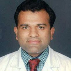
Dr Girish Velis
MBBS, DNB (Ophth”lmology), FAICO, FVRS, MNAMS
Consultant Ophthalmologist and Vitreoretina Surgeon

Dr Devika Dattatraya Lawande
FMR, FROP, DNB, Ophthalmology, D. O. M. S, M. B. B. S

Dr K”ushik U Dhume
MD ,DNB, MBBS


